When implementing tools for the diagnosis of multiple sclerosis, there are several aspects to take into account. The first one is that the patient may come to the clinic in a stable situation or with a flare-up and, in the case of the latter scenario, it may take time for the flare-up to recur.
This makes it necessary to have all resources available to gather the necessary information, detect the disease early and start treating it as soon as possible.
Diagnosing multiple sclerosis
In order to make a diagnosis of multiple sclerosis, it must be considered that the symptoms cannot always be clearly attributed to the disease. Coordination problems, slight changes in speech and weakness in the arms and legs, some of the symptoms of multiple sclerosis, can also appear in other pathologies that must be ruled out.
In addition, in order to make a reliable diagnosis, demyelinating lesions in at least two different neurological regions must be observed objectively.
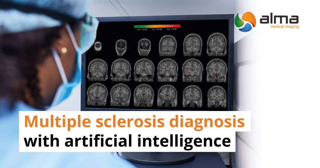
MRI detection
How can multiple sclerosis be detected?
Medical imaging plays a fundamental role in establishing a diagnosis, beyond the clinical observation of symptoms. In particular, we are talking about magnetic resonance imaging and especially its conventional sequences such as T1.
In fact, MRI has proven to be the most sensitive option for detecting demyelinating lesions in multiple sclerosis. It is therefore an excellent diagnostic test to differentiate these lesions from other lesions of the central nervous system.
Artificial intelligence for the diagnosis of multiple sclerosis
We can go one step further. As we have discussed in previous articles, artificial intelligence for medical diagnosis is a very good way to eliminate the inter-observer difference and make an objective and reliable assessment of the patient.
Below we list the tools that Qubiotech brings to our marketplace for the detection of this disease:
- Identification of atrophic VOI in MRI T1 sequences to have quantitative information about grey and white matter atrophies in different volumes of interest.
- Detection of hypermetabolic and hypometabolic areas from the deviation from the normal base in MRI, CT and PET images.
- Calculation of SUVR value, with PET and MRI, and MRI segmentation of grey matter, white matter and cerebrospinal fluid.
To discover our tools for the diagnosis of multiple sclerosis, enter our artificial intelligence for medicine marketplace and contact us to solve your doubts.